Description
Color–Switch (Green to Red), CRE reporter stable cell line:
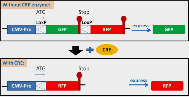
This cell line was transformed from HEK293 Cell line, expressing “LoxP-GFP-stop-LoxP-RFP” cassette under CMV promoter (see Color-Switch cassette below). Cell line also contains a Blasticidin antibiotic marker under RSV promoter (not shown in the scheme).
Applications:
- This CRE reporting cell lines is used to monitor the efficiency of CRE recombination in vivo. It is a great method and easy tool to verify the recombination event by your CRE enzyme (your CRE expression plasmids, or pre-made CRE expression lentivirus, or purified CRE protein) In vivo and In vitro .
- It is a convenient tool to verify your CRE-loxP based system.
- It can also used to verify your CRISPR editing efficiency by targeting at LoxPs and GFP/RFP region (please be advised, we do not provide any sequence information unless a license is purchased for this cell line).
How it works?
The cell line demonstrates strong GFP (Green Fluorescent signal). The downstream RFP (Red Fluorescent Protein) was not expressed because of the stop codon after the GFP. Once the CRE protein is present in nuclear, the CRE excises / deletes the DNA fragment between two loxP sites, which removes the stop codon (see the DNA structure scheme above). As a result, the RFP is then expressed, and the cell line switches to RFP fluorescent. The GFP and RFP signal can be easily monitored via fluorescent cell sorting (for the ratio between GFP and RFP cells), or visualized under Fluorescent microscope, or measured the fluorescent intensity if desired.
Sample images of CRE-loxP recombination detection:
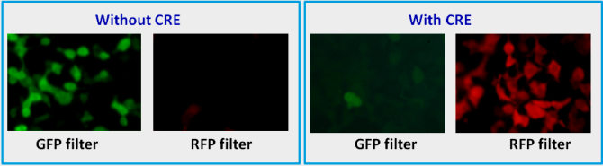
Left panel / without CRE: CRE reporter cell line (Cat#: SC018-Bsd) was cultured in completed. Images were taken under microscope with GFP filter set (Ex 490nm/Em 525nm) and RFP filter set (Ex 545nm/Em 620nm).
Right panel / with CRE: CRE reporter cell line was cultured in completed in 24-well plate. 50ul of CRE expression lentiviral particle (Cat#: LVP339) was added into the cells in one well. Images were taken at ~ 72 hours after the addition of CRE expression lentivirus.
Please click here for Product Manual.
Sold at: 1 vial x (2 x 10⁶ cells)/vial
Cat#: SC018-Bsd


