Cytoskeleton Imaging
Live Cell Imaging of Cytoskeleton Structure:
A fluorescent marker (GFP, RFP or CFP) fusion with a cellular structure protein, provides a convenient tool for visualization of cytoskeletal structure and dynamics under a variety of physiological and pathological treatment conditions, in live or fixed tissues and cells.
About Cell Skeleton imaging Lentivirus:
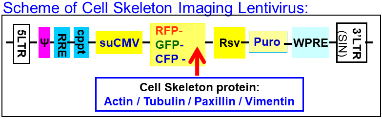
Utilizing Gentarget’s proprietary lentivector system, A fluorescent marker tagged an endogenous cell skeleton structure protein, and expressed under an enhanced CMV promoter. Depending upon the tagged structure protein, those lentivirus products are used to visualize microfilaments (β-actin), microtubules (α-tubulin), focal adhesions (paxillin) and intermediate filaments (vimentin). Under Fluorescent-microscope, the cytoskeletal structure demonstrated a fluorescent signal. It provides a convenient method for visualization of cytoskeletal structure and dynamics in real-time.
The lentivirus also contains a Puromycin antibiotic selection. The core structure of the lentivirus is shown at the following scheme.